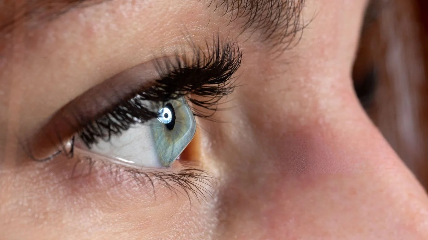
Keratoconus is a progressive eye condition that affects the cornea, the transparent, dome-shaped layer at the front of the eye. In individuals with keratoconus, the normally round cornea becomes thin and bulges into a cone-like shape. This change in shape distorts vision and can lead to significant visual impairment if left untreated. The condition typically begins in the teenage years or early adulthood and can worsen over time, but with early diagnosis and appropriate treatment, the progression can be slowed, and symptoms managed.
This article will explore the causes, symptoms, diagnosis, and treatment options for keratoconus, providing a comprehensive guide to understanding and managing this condition.
What is Keratoconus?
The cornea plays an essential role in focusing light onto the retina, allowing us to see clearly. In keratoconus, the cornea weakens and thins, causing it to bulge out into a cone shape instead of maintaining its usual rounded curvature. As a result, the shape of the cornea changes, leading to blurred or distorted vision. The condition typically affects both eyes, though one eye may be more severely affected than the other.
Keratoconus is classified as a progressive disease, meaning that its symptoms tend to worsen over time. If left untreated, it can lead to severe vision impairment, but with proper care, the condition can be managed effectively.
Causes of Keratoconus
The exact cause of keratoconus remains unclear, but several factors are thought to contribute to its development:
- Genetic Factors: Keratoconus tends to run in families, suggesting a genetic predisposition. Individuals with a family history of keratoconus are at a higher risk of developing the condition.
- Hormonal Changes: Keratoconus often begins during adolescence or early adulthood, a time when significant hormonal changes occur. Hormonal fluctuations, particularly during puberty, may play a role in the development and progression of the condition.
- Environmental Factors: Certain environmental factors, such as frequent eye rubbing, may contribute to the progression of keratoconus. Rubbing the eyes can increase stress on the cornea and exacerbate the condition.
- Other Health Conditions: Keratoconus is sometimes associated with other medical conditions, including Down syndrome, Ehlers-Danlos syndrome, and other connective tissue disorders.
Symptoms of Keratoconus
The symptoms of keratoconus vary depending on the severity of the condition and how far it has progressed. Early symptoms may be mild, but over time, the condition can cause significant vision problems. Common symptoms of keratoconus include:
- Blurred or Distorted Vision: Vision may appear blurry, especially for objects at a distance. The distortion can become more pronounced as the condition progresses.
- Increased Sensitivity to Light: People with keratoconus may develop photophobia, or increased sensitivity to bright light, making it difficult to see in well-lit environments.
- Frequent Changes in Prescription: Individuals with keratoconus often experience frequent changes in their eyeglass or contact lens prescriptions, as their vision continues to deteriorate.
- Halos and Ghosting: Affected individuals may see halos or ghost-like images around lights, particularly at night, due to the irregular curvature of the cornea.
- Eye Strain and Discomfort: The visual distortions caused by keratoconus may lead to eye strain, headaches, and discomfort, particularly when reading or using a computer.
Diagnosis of Keratoconus
Keratoconus can be diagnosed through a comprehensive eye exam. The eye doctor will typically perform a series of tests to assess the cornea’s shape, thickness, and the overall health of the eyes:
- Corneal Topography: This is the most important test for diagnosing keratoconus. It creates a detailed map of the cornea’s shape, showing any irregularities or bulging. This test can detect the condition early, even before noticeable changes in vision occur.
- Slit-Lamp Examination: A slit-lamp exam allows the eye doctor to examine the cornea under magnification, looking for signs of thinning or distortion.
- Pachymetry: This test measures the thickness of the cornea. People with keratoconus typically have a thinner cornea than normal.
- Keratometry: This test measures the curvature of the cornea, which may be more steeply curved in individuals with keratoconus.
Treatment Options for Keratoconus
While there is no cure for keratoconus, there are several treatment options available to manage the condition and slow its progression. The choice of treatment depends on the severity of the disease and the individual’s needs.
1. Glasses and Contact Lenses
In the early stages of keratoconus, vision can often be corrected with glasses or soft contact lenses. However, as the condition progresses and the cornea becomes more irregular, the following options may be recommended:
- Rigid Gas Permeable (RGP) Contact Lenses: These lenses are often the first choice for people with keratoconus. RGP lenses provide a smooth surface over the irregular cornea, improving vision. They may require frequent adjustments as the condition changes.
- Scleral Lenses: For more advanced cases of keratoconus, scleral lenses are large-diameter lenses that rest on the sclera (the white part of the eye) and create a reservoir of fluid over the cornea. These lenses provide better comfort and vision for individuals with severe keratoconus.
- Hybrid Lenses: Hybrid lenses combine a rigid center for better vision and a soft outer ring for comfort. They are suitable for individuals who need the clarity of RGP lenses but find them uncomfortable.
2. Corneal Cross-Linking (CXL)
Corneal cross-linking is a relatively new procedure that strengthens the cornea and helps to halt the progression of keratoconus. It involves applying riboflavin (vitamin B2) drops to the cornea and exposing it to ultraviolet (UV) light. This process increases the collagen fibers in the cornea, making the cornea stiffer and less prone to further bulging. Corneal cross-linking is most effective when performed early in the disease’s progression.
3. Intacs (Corneal Implants)
Intacs are small, ring-shaped devices that are inserted into the cornea to reshape it and reduce its bulging. The implants help to flatten the cornea, improving vision and reducing the severity of the condition. Intacs are typically used in moderate cases of keratoconus when other treatments have not been effective.
4. Corneal Transplant
In severe cases of keratoconus, when other treatments are no longer effective, a corneal transplant may be necessary. There are two main types of corneal transplants:
- Penetrating Keratoplasty (PK): This procedure involves replacing the entire cornea with a donor cornea.
- Lamellar Keratoplasty (LK): In this procedure, only the damaged layers of the cornea are replaced, leaving the healthy layers intact. This method is often preferred as it reduces the risk of complications.
5. Collagen Cross-Linking with Topography-Guided Photorefractive Keratectomy (PRK)
For individuals with moderate to advanced keratoconus, a combination of corneal cross-linking and PRK may be used to improve both the shape of the cornea and its stability. PRK involves the use of a laser to reshape the cornea, and when combined with cross-linking, it can offer significant improvement in vision and stability.
Preventing Keratoconus
Since the exact cause of keratoconus is unknown, there are no guaranteed ways to prevent it. However, some steps may reduce the risk of progression:
- Avoid Eye Rubbing: Rubbing the eyes can exacerbate keratoconus, so it’s important to avoid the habit of eye rubbing, particularly in individuals with a family history of the condition.
- Regular Eye Exams: Early detection through regular eye exams can help detect keratoconus in its early stages, allowing for prompt treatment and better management of the condition.
- Protective Eyewear: Wearing protective eyewear during physical activities can prevent eye injuries that may contribute to corneal damage.
Conclusion
Keratoconus is a progressive condition that affects the cornea and can lead to significant visual impairment if not treated. Although there is no cure, various treatment options are available to help manage the disease and improve vision, including glasses, contact lenses, corneal cross-linking, and in more severe cases, corneal transplants. Early diagnosis and appropriate treatment are key to managing keratoconus effectively and preventing further damage to the cornea. If you experience symptoms of keratoconus, such as blurred or distorted vision, it is important to seek professional care from an eye doctor for proper evaluation and treatment.
Frequently Asked Questions (FAQs)
Keratoconus is a progressive eye condition in which the cornea, the clear, dome-shaped outer layer of the eye, becomes thin and begins to bulge outward into a cone shape. This abnormal shape distorts the way light enters the eye, leading to blurry or distorted vision.
The exact cause of keratoconus is not fully understood, but several factors may contribute:
– Genetics: Keratoconus tends to run in families, suggesting a genetic predisposition.
– Hormonal changes: Keratoconus often develops during puberty or early adulthood, suggesting that hormonal changes may play a role.
– Eye rubbing: Chronic eye rubbing can aggravate the condition and lead to worsening of the corneal distortion.
– Other health conditions: Keratoconus is sometimes associated with conditions like Down syndrome, Ehlers-Danlos syndrome, or other connective tissue disorders.
Common symptoms of keratoconus include:
– Blurred or distorted vision, especially at night
– Frequent changes in prescription for glasses or contact lenses
– Increased sensitivity to light (photophobia)
– Ghosting or halos around lights
– Eye strain, discomfort, or redness
The symptoms often worsen as the condition progresses, and vision can become more difficult to correct with glasses alone.
Keratoconus is diagnosed through a comprehensive eye exam, including:
– Corneal topography: This test maps the surface of the cornea to detect irregularities and bulging.
– Slit-lamp examination: The eye doctor uses a specialized microscope to examine the cornea for signs of thinning or distortion.
– Pachymetry: This test measures the thickness of the cornea.
– Keratometry: Measures the curvature of the cornea to detect steepening, which is characteristic of keratoconus.
Currently, there is no known way to prevent keratoconus. However, avoiding habits such as chronic eye rubbing may reduce the risk of worsening the condition. Early diagnosis and treatment can help manage the disease and slow its progression.
Treatment options for keratoconus depend on the severity of the condition:
– Eyeglasses or soft contact lenses: In the early stages, glasses or soft contact lenses may correct vision.
– Rigid gas permeable (RGP) contact lenses: These lenses are commonly used to improve vision in individuals with keratoconus by smoothing the irregularities of the cornea.
– Corneal cross-linking (CXL): This procedure strengthens the cornea by using ultraviolet light and riboflavin (vitamin B2) to create collagen bonds in the cornea, preventing further thinning and bulging.
– Intacs: These are small, ring-shaped implants inserted into the cornea to reshape it and improve vision.
– Corneal transplant: In severe cases, a corneal transplant may be necessary if other treatments do not provide adequate results.
Corneal cross-linking (CXL) is a minimally invasive treatment that strengthens the cornea by applying riboflavin drops to the eye and exposing it to ultraviolet (UV) light. This process creates stronger collagen bonds in the cornea, helping to stabilize its shape and prevent further progression of keratoconus. CXL is often effective when performed early in the condition’s progression.
Intacs are small, curved inserts that are placed into the cornea to flatten and reshape it, improving its overall shape and helping to correct vision. These inserts do not replace the cornea but work to reduce the bulging and improve the eye’s focusing ability. Intacs are typically used in cases where keratoconus is moderate and when contact lenses are not effective.
In the early stages of keratoconus, glasses may still help correct vision. However, as the condition progresses and the cornea becomes more irregular, glasses may become less effective. Rigid gas permeable (RGP) contact lenses are commonly prescribed for individuals with keratoconus, as they provide better vision correction by creating a smooth surface over the cornea.
A corneal transplant may be necessary in advanced cases of keratoconus when other treatments, such as contact lenses or corneal cross-linking, are no longer effective in managing the condition. A corneal transplant involves replacing the damaged cornea with a healthy donor cornea and is typically considered when vision is severely impaired and cannot be corrected by other means.
While keratoconus can cause significant vision problems, the condition itself typically does not lead to complete blindness. With proper management, including the use of corrective lenses, corneal cross-linking, or a corneal transplant, many people with keratoconus can maintain functional vision. However, without treatment, the condition can lead to severe visual impairment.
The prognosis for people with keratoconus varies. In the early stages, the condition can be managed effectively with glasses or contact lenses. As the disease progresses, treatments like corneal cross-linking or Intacs can stabilize the cornea and improve vision. In severe cases, a corneal transplant may be required. With early intervention and proper treatment, many individuals with keratoconus can lead normal, active lives.
Regular eye exams are essential for people with keratoconus, as the condition can change over time. It is recommended to see an eye doctor at least once a year for monitoring, and more frequently if there are significant changes in vision or if the condition progresses. Your eye doctor will help determine the appropriate treatment plan and make any necessary adjustments.
While keratoconus typically affects both eyes, it can develop in one eye first and then progress to the other eye over time. It is important to monitor both eyes regularly for any signs of changes in vision.
Yes, keratoconus often runs in families, suggesting a genetic component to the condition. If you have a family history of keratoconus, you may be at higher risk of developing the condition yourself. Early detection is important, so individuals with a family history should undergo regular eye exams.
Leave A Comment